Venn diagram of meiosis and mitosis – Venn diagrams are an effective tool for visualizing and comparing the similarities and differences between two processes. In the context of cell division, venn diagrams can be used to compare meiosis and mitosis. These two processes are essential for the growth, development, and reproduction of all living organisms.
In this article, we will explore the venn diagram of meiosis and mitosis, examining the key stages, chromosome behavior, and outcomes of each process.
Comparison of Meiosis and Mitosis

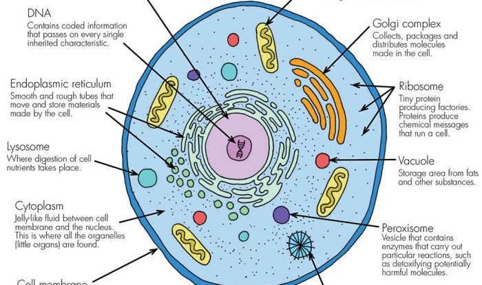
Meiosis and mitosis are two distinct cell division processes that occur in eukaryotic cells. Mitosis is the process by which a single cell divides into two identical daughter cells, while meiosis is the process by which a single cell divides into four haploid daughter cells.
Both mitosis and meiosis involve a series of stages, including prophase, metaphase, anaphase, and telophase. However, there are some key differences between the stages of mitosis and meiosis.
Stages of Mitosis and Meiosis
The following table summarizes the key differences between the stages of mitosis and meiosis:
| Stage | Mitosis | Meiosis |
|---|---|---|
| Prophase | Chromosomes condense and become visible. The nuclear envelope breaks down. | Chromosomes condense and become visible. The nuclear envelope breaks down. Homologous chromosomes pair up and exchange genetic material through a process called crossing over. |
| Metaphase | Chromosomes line up in the center of the cell. | Homologous chromosomes line up in the center of the cell. |
| Anaphase | Sister chromatids separate and move to opposite poles of the cell. | Homologous chromosomes separate and move to opposite poles of the cell. |
| Telophase | Two new nuclear envelopes form around the daughter chromosomes. The cell membrane pinches in the middle, dividing the cell into two daughter cells. | Four new nuclear envelopes form around the daughter chromosomes. The cell membrane pinches in twice, dividing the cell into four daughter cells. |
As you can see, the stages of mitosis and meiosis are very similar. However, there are some key differences that distinguish the two processes.
Chromosome Behavior

In meiosis, homologous chromosomes pair up and exchange genetic material through a process called crossing-over. These pairs of chromosomes, known as tetrads, then separate during meiosis I, resulting in two daughter cells with half the number of chromosomes as the parent cell.
During meiosis II, the sister chromatids of each chromosome separate, resulting in four daughter cells with half the number of chromosomes and half the amount of genetic material as the parent cell.
In mitosis, on the other hand, chromosomes do not pair up or exchange genetic material. Instead, each chromosome is duplicated, and the sister chromatids are separated during mitosis, resulting in two daughter cells with the same number of chromosomes and the same amount of genetic material as the parent cell.
Pairing of Chromosomes
In meiosis, homologous chromosomes pair up during prophase I through a process called synapsis. The homologous chromosomes are then held together by a protein complex called the synaptonemal complex. During this time, genetic material is exchanged between the homologous chromosomes through a process called crossing-over.
This exchange of genetic material results in the formation of new chromosomes that contain a combination of genetic material from both parents.
In mitosis, chromosomes do not pair up. Instead, each chromosome is duplicated during interphase, and the sister chromatids are held together by a protein complex called cohesin. The sister chromatids are then separated during mitosis, resulting in two daughter cells with the same number of chromosomes and the same amount of genetic material as the parent cell.
Separation of Chromosomes
In meiosis, the homologous chromosomes separate during meiosis I, and the sister chromatids separate during meiosis II. This separation of chromosomes results in the formation of four daughter cells with half the number of chromosomes and half the amount of genetic material as the parent cell.
In mitosis, the sister chromatids separate during mitosis, resulting in two daughter cells with the same number of chromosomes and the same amount of genetic material as the parent cell.
Number of Daughter Cells: Venn Diagram Of Meiosis And Mitosis

Meiosis and mitosis differ significantly in the number of daughter cells they produce. Meiosis, a process of cell division that occurs in reproductive cells, results in the formation of four genetically distinct daughter cells, known as gametes (eggs or sperm).
In contrast, mitosis, a process of cell division that occurs in somatic cells (body cells), results in the formation of two genetically identical daughter cells. This difference in the number of daughter cells produced by meiosis and mitosis is crucial for maintaining the appropriate chromosome number in organisms.
Significance of Different Daughter Cell Numbers
The different numbers of daughter cells produced by meiosis and mitosis have significant implications for the genetic diversity and continuity of organisms:
- Meiosis:The production of four genetically distinct daughter cells through meiosis ensures genetic variation in offspring. This genetic diversity is essential for adaptation and survival in changing environments.
- Mitosis:The production of two genetically identical daughter cells through mitosis ensures the faithful transmission of genetic information during cell growth, tissue repair, and asexual reproduction. It maintains the genetic stability and continuity of the organism.
Genetic Variation

Meiosis plays a crucial role in genetic variation, the process that generates diversity within a species. This variation is essential for adaptation, evolution, and the survival of species in changing environments.
Genetic variation arises during meiosis through two primary mechanisms: crossing over and independent assortment.
Crossing Over
Crossing over occurs during prophase I of meiosis, when homologous chromosomes pair up and exchange genetic material. This process results in the formation of recombinant chromosomes, which contain a unique combination of alleles from both parents.
Independent Assortment, Venn diagram of meiosis and mitosis
Independent assortment refers to the random distribution of chromosomes during meiosis II. Each gamete receives a random assortment of maternal and paternal chromosomes, ensuring that each gamete contains a unique combination of alleles.
The combination of crossing over and independent assortment generates a vast array of possible gamete combinations, each with a unique genetic makeup. This genetic diversity provides the raw material for natural selection to act upon, driving the evolution of species over time.
FAQ Resource
What is the main difference between meiosis and mitosis?
The main difference between meiosis and mitosis is the number of daughter cells produced. Meiosis produces four daughter cells, while mitosis produces two daughter cells.
What is the role of meiosis in genetic variation?
Meiosis plays a crucial role in genetic variation by creating gametes (eggs and sperm) with unique combinations of chromosomes. This variation is essential for the survival and evolution of species.
What are the stages of meiosis?
The stages of meiosis are prophase I, metaphase I, anaphase I, telophase I, prophase II, metaphase II, anaphase II, and telophase II.